What is the Carotid Artery?
Carotid stenosis is a narrowing of the carotid arteries, the two main arteries that carry oxygen-rich blood from the heart to the brain. Carotid artery stenosis, also called carotid artery disease, is caused by a buildup of plaque (atherosclerosis) in the artery wall that reduces blood flow to the brain. Treatment aims to reduce the risk of stroke by controlling or removing plaque buildup.
Blood Circulation of the Brain
To understand carotid artery stenosis, it is first necessary to understand the circulatory system of the head and neck. The carotid artery, also known as the carotid artery, originates from the aorta in the chest and travels from the neck to the head. When you place your hands on either side of your neck, you can feel your pulse in your carotid arteries. Near the larynx (Adam’s apple), the carotid artery divides into the external and internal carotid arteries. The external artery supplies blood to the face and scalp, while the internal artery supplies blood to the brain. The most common site of atherosclerotic plaque accumulation is the carotid bifurcation, where the carotid artery divides into internal and external branches.
What is Carotid Artery Stenosis?
Carotid stenosis is the gradual narrowing of the carotid arteries, a process called atherosclerosis. Normal, healthy arteries are flexible and have smooth inner walls. As we age, hypertension and minor injuries to the blood vessel wall can lead to plaque formation. Plaque is a sticky substance made of fat, cholesterol, calcium, and other fibrous material. Over time, plaque deposits on the inner wall of the artery can form a large mass that narrows the lumen, the inner diameter of the artery. Atherosclerosis also causes the arteries to harden, often called “hardening of the arteries.”
There are three ways that carotid artery stenosis increases the risk of stroke:
- Plaque deposits grow larger, severely narrowing the artery and reducing blood flow to the brain. Eventually, the plaque can completely block the artery.
- Plaque deposits can roughen and deform the inner surface of the artery. This causes blood clots to form inside the artery, blocking blood flow to the brain.
- Plaque deposits can rupture or break. The ruptured or broken pieces travel through the artery toward the brain, blocking small blood vessels and preventing blood flow to the brain.
What are the symptoms?
Most people with carotid artery stenosis have no symptoms until the artery is severely narrowed or a clot forms. Symptoms are most likely to first appear in a mini-stroke, also known as a transient ischemic attack (TIA). TIAs occur when blood flow to the brain is temporarily interrupted and stops when it starts again. Symptoms typically last for a few minutes and then completely resolve and the person returns to normal. TIAs should never be ignored; they are early warning signs of an ischemic stroke and permanent brain damage. Symptoms of a TIA or ischemic stroke can include weakness or numbness in an arm or leg, difficulty speaking, facial squinting, vision problems, or paralysis affecting one side of the body. If you or a loved one experiences these symptoms, you should call 911 immediately.
What are the reasons?
Atherosclerosis is the main cause of carotid artery disease. It can begin in adolescence, but it takes decades for symptoms to appear. For some people, atherosclerosis progresses rapidly in their thirties, while for others it can occur in their fifties or sixties. Atherosclerosis damages the inner lining of the arteries due to high blood pressure, diabetes, smoking, and high cholesterol—especially the “bad” cholesterol, or low-density lipoprotein (LDL). Other risk factors include obesity, coronary artery disease, a family history of carotid artery stenosis, and older age.
Less commonly, carotid aneurysm and fibromuscular dysplasia can also cause carotid stenosis.
People with heart disease are at higher risk of developing carotid artery stenosis. Typically, these arteries become diseased several years later than the coronary arteries.
Who is at Risk?
Older people are more likely to be affected by carotid artery stenosis. Before the age of 75, men are at greater risk than women. A person with high cholesterol, high blood pressure and who smokes is eight times more likely to develop atherosclerosis than a person without risk factors. More than 500,000 new strokes occur each year in the United States, and carotid artery stenosis is estimated to cause 20% to 30% of these strokes.
How is Diagnosis Made?
Your doctor will ask you some questions to learn as much as possible about your symptoms, medical problems, medications you are taking, and family history. Then, he or she will examine you. Your doctor may listen to your carotid artery with a stethoscope to hear a bruit. If a bruit is heard, it may be a sign of turbulent blood flow caused by atherosclerosis. In suspected patients, certain diagnostic tests, such as a Doppler ultrasound of the neck, CT angiogram (CTA) of the neck, magnetic resonance angiography (MRA), or cerebral angiography, are performed to definitively detect narrowing of the carotid artery.
Imaging methods such as MRI and CT can also reveal stroke foci in the brain. When tests show reduced blood flow in one or both carotid arteries, your doctor will diagnose carotid stenosis.
- A Doppler ultrasound is a simple test that uses reflected sound waves to assess blood flow through your arteries. The ultrasound probe is placed in your neck area above your carotid artery. This test will reveal how much blood is flowing through the artery and the degree to which the artery is narrowed (i.e. 100%, 80%, 70%, etc.).
- Computed Tomography Angiography, or CT angiography, is a noninvasive X-ray that provides detailed images of anatomical structures in the brain. It involves injecting a contrast material into the bloodstream to see the arteries of the brain. This type of test provides the best images of both blood vessels (via angiography) and soft tissues (via CT). It allows your doctor to see the narrowed artery and determine how narrowed it is.
- A Magnetic Resonance Angiography (MRA) of the neck is similar to a CT angiogram. Contrast dye is injected through a vein to illuminate the blood vessels in the neck.
- Cerebral Angiogram is an invasive test that involves inserting a catheter into the main artery in the groin and advancing it to the exit areas of the carotid arteries, where the contrast material injected into these areas becomes visible with the help of X-rays. This method allows your doctor to see your carotid arteries and all the arteries in your brain.
What Treatment Methods Are Available?
The goal of treatment is to reduce the risk of stroke. Treatment options depend on the severity of the narrowing of the carotid artery and whether you are experiencing stroke-like symptoms.
Drug Therapy
People with no symptoms or less than 50% carotid artery stenosis are usually treated with medication. People with a medical condition that increases their risk of surgery may also be treated with medication. Medications include:
- Antiplatelet drugs (aspirin, ticlopidine, clopidogrel) thin the blood and prevent clotting in narrowed arteries, allowing blood to flow through the vessel more easily.
- Cholesterol drugs help reduce plaque formation in atherosclerosis. These drugs, called statins, can reduce LDL “bad” cholesterol by an average of 25-30% when combined with a low-fat diet.
- Antihypertensive drugs (diuretics, ACE inhibitors, angiotensin blockers, beta blockers, calcium channel blockers, etc.) help control and regulate blood pressure. Regular blood pressure screenings are recommended because high blood pressure poses a serious risk of stroke.
Surgical Treatment
Surgery is usually recommended for patients who have had one or more TIAs or strokes and have moderate to severe carotid artery stenosis. The goal of surgery is to remove plaque and widen the artery walls to allow more blood to flow to the brain, preventing a stroke. The methods used include:
Carotid Endarterectomy

This is open surgery to remove plaque. A skin incision is made in the neck and the carotid artery is found. Temporary clamps are placed in the artery areas above and below the narrowing area to stop blood flow. During this time, the carotid artery on the other side of the neck continues to supply blood to the entire brain. The surgeon makes an incision in the artery over the blocked area. The plaque buildup is peeled away and removed (Figure 1). You can watch our videos here that show in detail how these surgeries are performed.

The artery is then closed with fine sutures, and the clamps are removed to restore bilateral blood flow to the brain (Figure 2). Carotid endarterectomy is typically indicated for patients with symptoms (stroke or TIA) and a greater than 50% blockage. It is also recommended for patients with no symptoms and a greater than 60% blockage. Surgery for patients with moderate blockage of 50 to 69% reduces the risk of stroke by 6.5% over a five-year period. Among patients with high-grade blockage of more than 70%, the risk of stroke is reduced by 80%. In patients with a stenosis of 50% or less, the benefits of endarterectomy do not outweigh the risks of surgery.
Carotid Artery Angioplasty (Balloon) and Stenting
This is an invasive procedure that compresses plaque and widens the inside of the artery. It is performed with an angiography device in the radiology suite. A flexible catheter is inserted into the groin artery. It is guided through the blood flow through the heart and into the carotid artery. A small catheter with an inflatable balloon on its tip is then placed over the narrowing plaque.
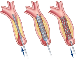

The balloon is expanded to widen the artery and compress the plaque against the artery wall. The balloon is then deflated and removed. Finally, a self-expanding, mesh-like metal cage called a stent is placed over the plaque to open the artery (Figure 3). Angioplasty (balloon) and stenting are generally reserved for select patients who: 1) have moderate to severe carotid artery stenosis of greater than 70%; 2) have other medical conditions that increase the risk of surgical complications; 3) have recurrent stenosis; or 4) have stenosis caused by radiation therapy to the neck for cancer treatment.
Carotid Artery Bypass Surgery
This operation is a surgical treatment method performed on patients whose arteries are completely blocked by plaque. It is a method that allows the blockage to be overcome by stitching a vein taken from another part of the body, usually from the vein in the leg or the artery in the arm, below and above the blockage. Carotid artery bypass surgery is typically performed only in cases where the carotid artery is 100% blocked.
Healing and Protection
Depending on your risk factors, your doctor may want you to quit smoking, limit excessive alcohol consumption, maintain strict blood sugar control (if you have diabetes), have your cholesterol checked regularly, and take medications as prescribed.
After carotid endarterectomy, restenosis can occur in less than two years and is usually asymptomatic. These relapsed plaques can be treated with angioplasty (balloon) and stenting. The plaques may regress over time, and intervention is only indicated for stenosis greater than 80%. After two years, restenosis is strongly associated with progression of atherosclerotic disease. In general, repeat surgery or stenting is recommended for symptomatic restenosis or stenosis greater than 80%.
It is important to remember that carotid stenosis is a progressive disease. If left untreated, stroke occurs at a rate of 13% per year in people with symptoms of carotid stenosis and 2.2% per year in people without symptoms.